Xiralite United States
 Xiralite Deutschland
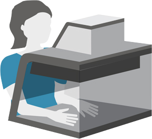
Imaging Workflow
Easy. Fast. Reliable.
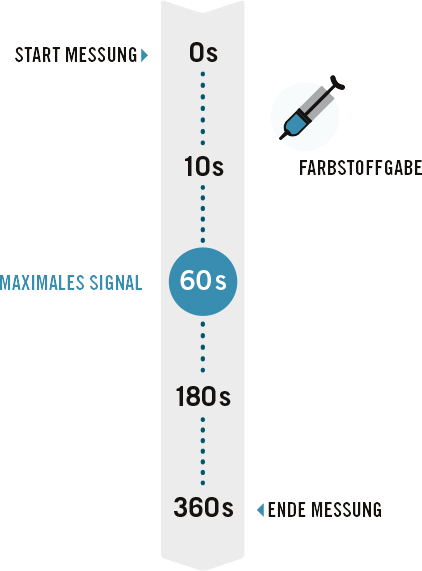
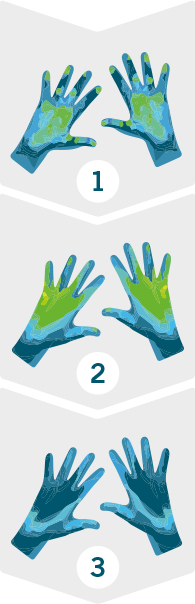
The Xiralite procedure allows a user-friendly imaging display with a high level of patient comfort:
- Free of radiation
- Fear- and stress free
- No exam preparation needed
- Short examination time (max. 10 minutes)
- Fast results
- High reliability