Xiralite United States
 Xiralite Deutschland
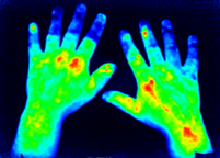
FOI (Xiralite) improves prediction of digital ulcers in systemic sclerosis using a composite score
Systemic sclerosis is a rare chronic disease characterized by diffuse fibrosis and vascular abnormalities in the skin, joints, and internal organs. In most patients, initial symptom is a blue or white coloring in response to cold temperature, a condition known as Raynaud’s phenomenon. Eventually, half of those persons will develop serious ischemic complications. They will get digital ulcers or pitting scars that quickly result in pain, loss of function or in the worst cases, amputation. Prediction of this outcome is vital in order to establish the right preventive treatment. Unfortunately, existing tools have a high sensitivity but a low specificity, meaning that some patients might receive treatments that they do not require.
We are glad to report that in a collaborative project involving several centers, a group of clinicians proved that fluorescence optical imaging (FOI – Xiralite) can improve diagnostic performance. FOI (see picture) was combined with doppler ultrasound, clinical data and patient history creating a so-called composite score (CIP-DUS). Following this score, predictions on who might develop digital ulcers and therefore need treatment, were higher.
These results were published in the Arthritis Research & Therapy journal and can be found at the following link.